This page contains data to support our paper on p53 mutation analysis.
Martin, A. C. R., Facchiano, A. M., Cuff, A. L., Hernandez-Boussard, T., Olivier, M., Hainaut, P. and Thornton, J. M. (2002) Integrating Mutation Data and Structural Analysis of the p53 Tumour-Suppressor Protein, Human Mutation, 19, 149-164. [PDF]
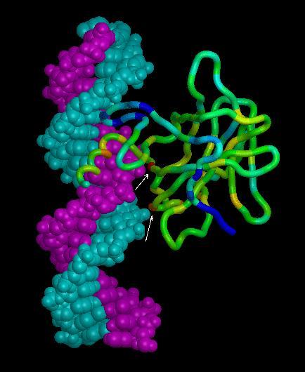
p53 is a nuclear phospho-protein which, in response to DNA damage, slows progression through the cell cycle and initiates apoptosis if damage is severe. Tumour-specific point mutations occur in many forms of human cancer with as many as 50% of cancers containing a p53 mutation. 20% of mutations are concentrated at 5 'hot-spot' codons. In order to understand mutations in the core domain in structural terms, a relational database has been developed containing mutation data, tumour details and results of structural analysis. An automated protocol has been developed using this database to attempt to explain the effects of mutations in structural terms.